What is Fluor-PSMA?
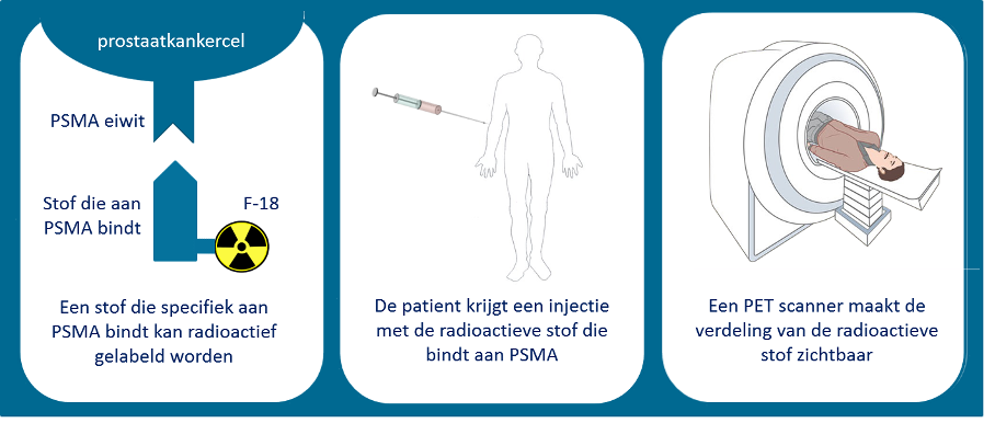
The radiopharmaceutical Fluor-18-PSMA is produced in a cyclotron. As mentioned on Gallium-PSMA, PSMA in the name does not refer to the protein in the cell membrane, but to the ligand that binds to the PSMA in the cell membrane. In a cyclotron, O-18 is irradiated with protons (H-1), from which the radioactive atom Fluor-18 is formed. This sets it apart from Gallium-PSMA, which is made in a laboratory in a hospital.
The production capacity of a cyclotron is many times higher than that of a Gallium generator. Additionally, the half-life of F-18 is approximately 110 minutes, making this radiopharmaceutical much more suitable for distribution to, and use in, surrounding centers.
Two types of Fluorine-PSMA are used in the Netherlands, namely: F-18-PSMA-1007 and F-18-DCFPyL. Both radiopharmaceuticals have a high affinity for PSMA in the cell membrane. However, the biological distribution of the two is different. This is partly due to the lipophilicity of PSMA molecules.
The distribution of F-18-DCFPyL in the body is like that of Ga-PSMA-11. Both tracers are primarily cleared through the kidneys resulting in a lot of radioactive urine in the urinary tract and bladder. This can have a disruptive effect on the scan, for example in the region of the prostate or seminal vesicles, which are both located directly below the bladder.
F-18-PSMA-1007 is highly lipophilic, which means it clears through the liver and bile ducts. There is little or no clearance via the kidneys, so that the local disturbance around the bladder and urinary tract is less than with PSMA-11 or DCFPyL. A recent article concludes that PSMA-1007 shows more non-specific lesions (non-tumor-related uptake) compared to PSMA-11, especially in bones.

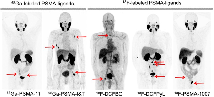
Figure 1. Several PSMA tracers in a row.
This figure shows scans with different tracers in different patients, with red arrows indicating where the prostate cancer metastases are located. This view provides insight into how the different tracers are distributed throughout the body. There are several visible differences that are important when assessing the scans. Usually we see normal (physiological) absorption in the salivary glands, lacrimal glands, liver (varying), kidneys and duodenum (the horizontally oriented piece of intestine located between the kidneys). DCFBC is the only one showing virtually no uptake in the lacrimal and salivary glands. However, there is relatively much activity in the vessels and the heart. This is also visible to a lesser extent at PSMA-I&T. A varying degree of uptake in the duodenum is often visible, PSMA-1007 also shows this in the other loops of the small intestine. PSMA-1007 is the only one to show minimal urinary and bladder excretion, while this is more pronounced in the first four tracers. Reference: PSMA Ligands for imaging and therapy. Eiber et al., JNM, Sept. 2017.


A nuclear medicine doctor evaluates a PET-CT scan.
